Blood Vessels Labeled ~ Cardiovascular System. The 4 valves are the aortic, pulmonary, mitral, and tricuspid valves. 15 minutes it would be impossible to get blood to the predestined locations without the vascular pathways. Blood vessels consist of arteries, arterioles, capillaries, venules, and veins. Free online quiz blood vessel labeling The smallest veins are called venules.
4 but is clearly visible entering the right atrium of the heart. The inferior vena cava is labeled in the figure below. Tunica intima—this is the inner thinnest layer. The smallest veins are called venules. Factors that affect blood pressure cardiovascular system:
Module 16 Blood Vessels Labeling Flashcards Quizlet from o.quizlet.com The eyeball is filled with vitreous humor, with the aqueous humor lying in the small anterior chamber of the eye. The function and structure of each segment of the peripheral vascular system vary depending on the organ it supplies. The left atrium receives blood from the lungs. They let blood flow forward and prevent the backward flow. Blood vessels consist of arteries, arterioles, capillaries, venules, and veins. Blood circulates throughout the body in blood vessels, propelled by the pumping action of the heart. Its smooth surface decreases resistance to blood flow Aside from capillaries, blood vessels are all made of three layers:
The common cartoid artery extends from the brachiocephalic artery.
Arteries and veins are composed of three tissue layers. Vessels transport nutrients to organs/tissues and to transport wastes away from organs/tissues in the blood. Blood supply to the scalp. The posterior auricular, occipital and superficial temporal arteries (along with two branches of the internal carotid artery; Aside from capillaries, blood vessels are all made of three layers: The venules and veins returning blood to the heart. The sclera and cornea (opaque and transparent layer respectively) the choroid (filled with blood vessels) Arteries (in red) are the blood vessels that deliver blood to the body. Interactive physiology with quizzes cardiovascular system: The iliac, femoral, popliteal and tibial (calf) veins are the deep veins in the legs. The common cartoid artery extends from the brachiocephalic artery. 4 but is clearly visible entering the right atrium of the heart. The eyeball is filled with vitreous humor, with the aqueous humor lying in the small anterior chamber of the eye.
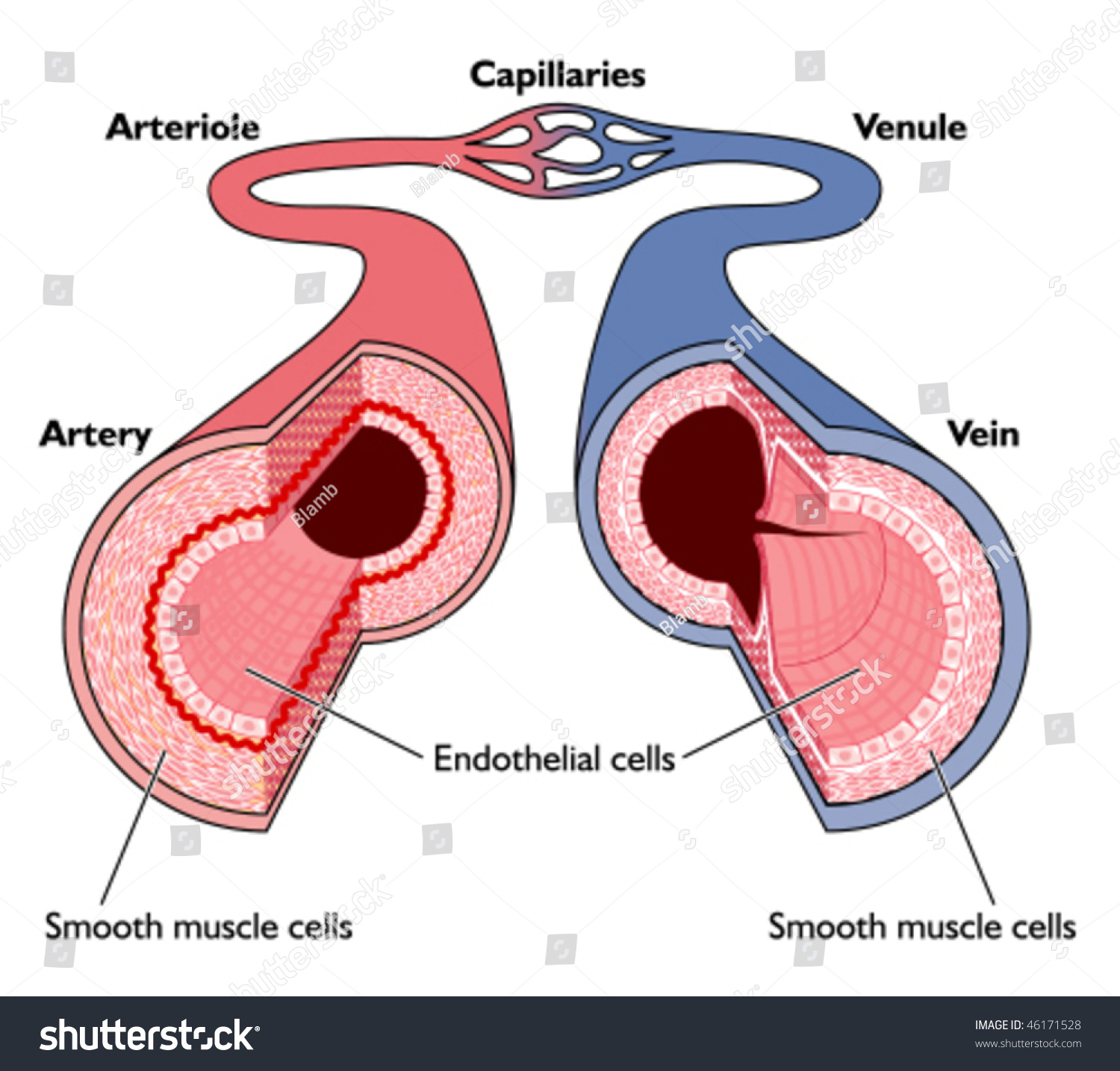
Blood vessels comprise the vascular system. Arteries carry blood away from the heart to other organs. Free online quiz blood vessel labeling The sclera and cornea (opaque and transparent layer respectively) the choroid (filled with blood vessels) Eventually, the smallest arteries, vessels called arterioles, further branch into tiny capillaries, where nutrients and wastes are exchanged, and then combine with other vessels that exit capillaries to form venules, small blood vessels that carry blood to a vein, a larger blood vessel that returns blood to the heart.
Anatomy Blood Vessels Artery Through Capillaries Stock Vector Royalty Free 46171528 from image.shutterstock.com This article lists a series of labeled imaging anatomy cases by system and modality. The superior vena cava is not labeled in figure 7.4. Arteries and veins are composed of three tissue layers. Blood supply to the scalp. The vessels that carry blood away from the heart are called arteries, and their very small branches are arterioles. Factors that affect blood pressure cardiovascular system: Blood vessels consist of arteries, arterioles, capillaries, venules, and veins. Start studying blood vessels labeling.
The superior vena cava is not labeled in figure 7.4.
Tunica intima—this is the inner thinnest layer. Blood vessels anatomy blood vessels are responsible for the transportation of blood, made up arteries and veins, they creates pathways for the oxygenated blood to travel to their destination and pathways for the used deoxygenated blood to travel back to the heart or lungs.capillaries are designed to permit the transfer of gasses within the blood, such as the delivery of oxygen and the return. A primary purpose and significant role of the vasculature is its participation in oxygenating the body. Blood vessels form a continuous path for blood flow that starts and ends at the heart.arteries carry blood away from the heart, regardless of the degree of blood oxygenation.veins carry blood toward the heart. The left atrium receives blood from the lungs. Free online quiz blood vessel labeling There are five main types of blood vessels: Blood vessel labeling online quiz; The function and structure of each segment of the peripheral vascular system vary depending on the organ it supplies. The sclera and cornea (opaque and transparent layer respectively) the choroid (filled with blood vessels) Blood vessels are found throughout the body. To play this quiz, please finish editing it. Like arteries, veins form a complex, branching system of larger and smaller vessels.
Blood vessels form the extensive networks by which blood leaves the heart to supply tissue. The videos are done by dr. Arteries and veins are composed of three tissue layers. The iliac, femoral, popliteal and tibial (calf) veins are the deep veins in the legs. They can vary in size.
Heart Anatomy Labeled Diagram Structures Blood Flow Function Of Cardiac System Ezmed from images.squarespace-cdn.com The videos are done by dr. The posterior auricular, occipital and superficial temporal arteries (along with two branches of the internal carotid artery; Interactive physiology with quizzes cardiovascular system: Best quiz blood vessel labeling; The three major types of blood vessels: The venules and veins returning blood to the heart. This video series covers the blood vessels for anatomy and physiology ii students. The sclera and cornea (opaque and transparent layer respectively) the choroid (filled with blood vessels)
The superior vena cava is not labeled in figure 7.4.
Additionally, other blood vessels return from. The common cartoid artery extends from the brachiocephalic artery. Very small branches that collect the blood from the various organs and parts are called venules, and they unite to form veins, which return the blood to the heart. The left atrium receives blood from the lungs. Arteries, veins, and capillaries blood vessels flow blood throughout the body. Aside from capillaries, blood vessels are all made of three layers: Blood vessels anatomy blood vessels are responsible for the transportation of blood, made up arteries and veins, they creates pathways for the oxygenated blood to travel to their destination and pathways for the used deoxygenated blood to travel back to the heart or lungs.capillaries are designed to permit the transfer of gasses within the blood, such as the delivery of oxygen and the return. Blood vessel labeling online quiz; The thick outermost layer of a vessel (tunica adventitia or tunica externa) is made of connective tissue. Blood vessels comprise the vascular system. The videos are done by dr. Tunica intima—this is the inner thinnest layer. This video series covers the blood vessels for anatomy and physiology ii students.